Learn About Our Products
Gallant is advancing veterinary care into the regenerative medicine era.
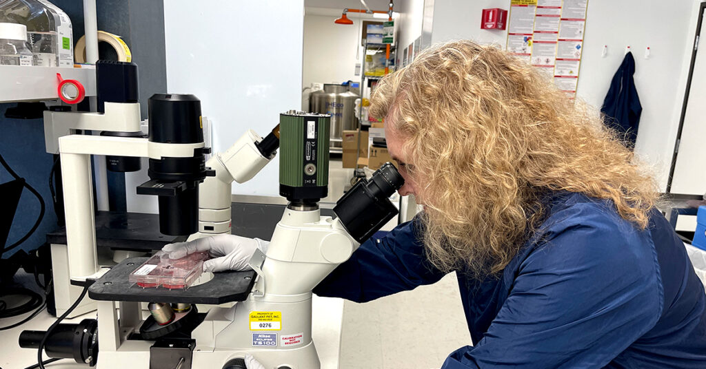
A New Category of Veterinary Medicine
We’re shaping a new category of treatment: ready-to-use cell therapies designed to address disease at its source rather than just manage symptoms.
Our investigational therapies are built on mesenchymal stromal (stem) cells (MSCs)—multipotent cells found in tissues such as bone marrow, uterus, and adipose. These cells help restore balance by influencing both innate and adaptive immunity, unlocking their unique regenerative potential.
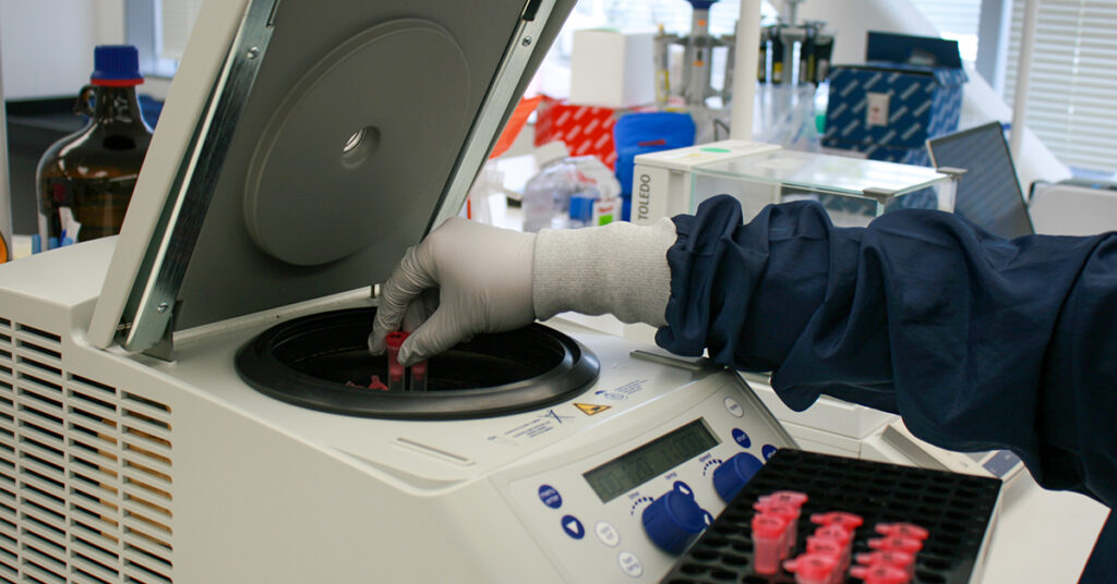
Science That Meets the Highest Standards
Every therapy in development follows FDA guidance for cell-based products. That means rigorous clinical trials, thorough safety evaluations, and strict manufacturing controls—all to ensure these therapies are both credible and clinically meaningful.
Disclaimer
Gallant’s investigational stem cell therapies are not commercially available. These veterinary products may be available through participation in a study at a qualified clinic under FDA-authorized protocols. This blog is intended for educational purposes only and does not constitute medical advice.